Alveolar bone expansion, also referred to as buccal bone expansion, is an interesting condition occurring primarily in middle-aged and older cats and occasionally, in dogs. It occurs with highest frequency around the maxillary canines, but it can also be seen around the mandibular canines, and rarely, lateral to the maxillary and mandibular premolars.
It is unknown why alveolar bone expansion occurs. It sometimes happens in areas of periodontal pocketing and vertical bone loss where bone is laid down laterally to buttress a periodontally unsound tooth (Figure 1). Alternatively, the addition of bone may be a reaction to the inflammation associated with periodontitis.
One study that assessed radiographic signs of periodontitis in cats found 53 percent of those in the study had expansion of alveolar bone at one or more canine tooth.1
A study published in 2015 evaluated alveolar bone biopsies submitted to pathologists. All samples showed clinical or radiographic evidence of periodontitis. Histologically, the tissue samples from canine tooth sites of cats had compressed trabeculae of mature remodeled bone, loose fibrous stroma with paucicellular inflammation, and mild proliferation of woven bone.2
Another study evaluated dental radiographs and electronic medical records of 97 cats. Results showed alveolar bone expansion was common in cats; breed, sex, and age predilections were not seen. Surprisingly, the study found no association between bone expansion, extrusion, and tooth resorption. A pattern of vertical alveolar bone loss is a typical feature of bone expansion, rather than horizontal bone loss.3
Alveolar bone expansion occasionally occurs in the absence of periodontitis. There are cases in which there is a marked bony proliferation around the tooth in the absence of gingival recession or periodontal pocketing (Figure 2).

The reasons for alveolar bone expansion in these cases are not known. Perhaps some are due to abnormalities in calciotropic hormones, such as 25-hydroxyvitamin D.
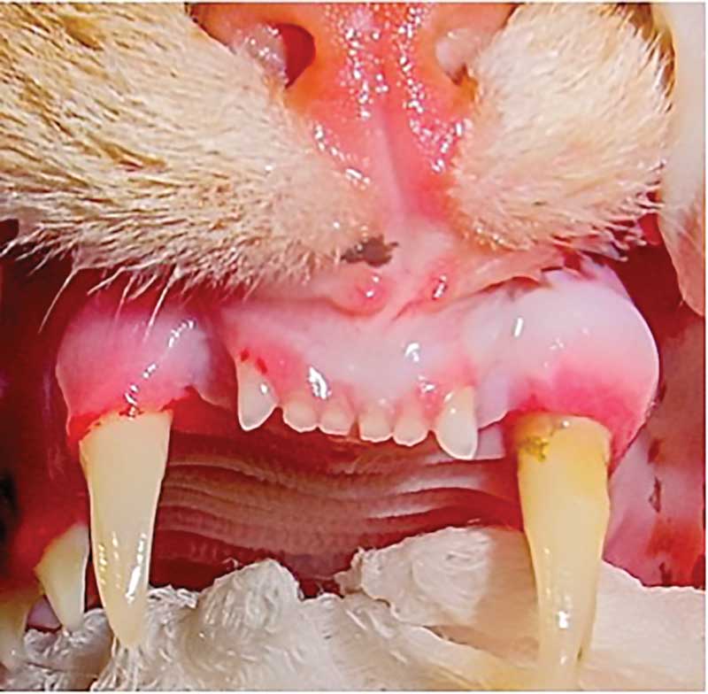
One study that looked at another oral disease of cats (tooth resorption) found a statistically significant increase in 25-hydroxyvitamin D in affected cats.4 Experimental over-supplementation of vitamin D and its metabolites in dogs and rats has revealed histological signs similar to those seen in cats, including hypercementosis and alveolar bone expansion (Figure 3).5 Cats are unable to produce vitamin D in their skin. Nutritional research has shown a plasma concentration of 25-hydroxyvitamin D in kittens is directly related to the dietary intake of vitamin D. It is possible alveolar bone expansion is a clinical manifestation of chronically increased serum levels of 25-hydroxyvitamin D levels.6


Prince, an approximately six-year-old male neutered shorthair cat, was presented recently with severe swelling of the rostral mandible. The swelling was so severe, that it would have been easy to blame it on something more insidious, such as cancer. The swelling was biopsied by Prince’s primary care veterinarian, which revealed alveolar bone expansion and osteomyelitis.
Prince was then referred to us to see if we agreed the clinical picture matched the histological findings. Oral examination showed a prominent, bilaterally symmetrical bony swelling of the rostral mandible (Figure 4 A). The swelling was so profound the maxillary canine teeth were impaling the soft tissue lateral to the bony swelling (Figure 4 B).
Other oral exam findings included missing teeth; multiple teeth affected by tooth resorption. Prince also showed alveolar bone expansion and extrusion of his maxillary canine teeth.


We placed Prince under anesthesia and raised a mucoperiosteal flap to expose the bone. Biopsies of the bone were collected with small rongeurs. The dog responded nicely to surgical reduction of excessive bone, extraction of the maxillary canine teeth that were impaling the area and removal of a retained root of the left mandibular canine tooth that may have been contributing to the osteomyelitis. All other teeth affected by tooth resorption and periodontal disease were extracted. Visual inspection of the mandibular bone in surgery showed no evidence or osteonecrosis. Prince was placed on a 10-day course of amoxicillin/clavulanate. Histopathology performed by a pathologist with a particular interest in oral pathology confirmed the previous diagnosis of alveolar expansion and osteomyelitis. Prince’s case is a good reminder that not all bony swellings are due to cancer. We are glad that Prince is doing well and has a good prognosis!
John Lewis, VMD, DAVDC, Fellow, AVDC OMFS, practices at Veterinary Dentistry Specialists and teaches at Silo Academy Education Center, both in Chadds Ford, Pa.
References
- Lommer MJ, Verstraete FJ. Radiographic patterns of periodontitis in cats: 147 cases (1998-1999). J Am Vet Med Assoc. 2001;218(2):230-234.
- Bell CM, Soukup JW. Histologic, Clinical, and Radiologic Findings of Alveolar Bone Expansion and Osteomyelitis of the Jaws in Cats. Vet Pathol. 2015;52(5):910-918.
- Peralta S, Fiani N, Scrivani PV. Prevalence, Radiographic, and Demographic Features of Buccal Bone Expansion in Cats: A Cross-Sectional Study at a Referral Institution. J Vet Dent. 2020;37(2):66-70.
- Reiter AM, Lyon KF, Nachreiner RF, Shofer FS. Evaluation of calciotropic hormones in cats with odontoclastic resorptive lesions. Am J Vet Res. 2005;66(8):1446-52.
- Reiter AM, Lewis JR, et al. Update on the etiology of tooth resorption in domestic cats. Vet Clin North Am Small Anim Pract 2005; 35: 913-942.
- Lewis JR, Okuda A, Shofer FS, Pachtinger G, Harvey CE, Reiter AM. Significant association between tooth extrusion and tooth resorption in domestic cats. J Vet Dent. 2008;25(2):86-95